
How to avoid the common mistakes when taking Dental Digital X-Rays
The intraoral dental x-ray is among the most powerful diagnostic weapons in the dentist’s arsenal. But after a while, it’s very easy to take x-rays for granted, to take sloppy shots, to make the same mistakes time and time again, and worse, unnecessarily expose patients to more radiation, as a direct consequence of retakes.
In this article we hope to inform you how you can minimize patient and operator exposure identify and proper errors in digital intraoral radiographs; how you can manage patients to obtain better shots and altogether improve the caliber of your radiography.
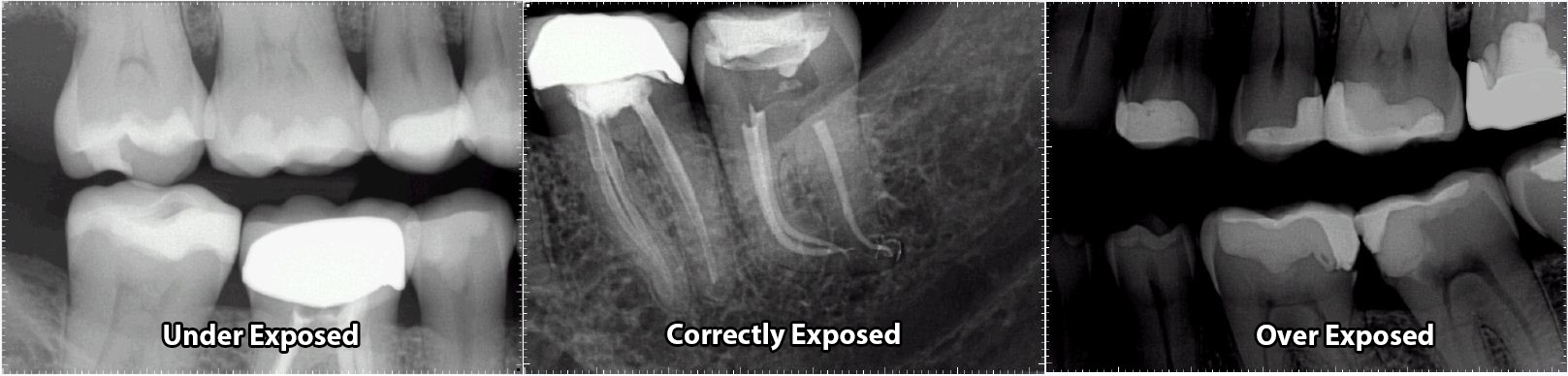
#1 Under/Over Exposure
The number one reason for poor radiographs…Exposure. Either your x-rays are coming out to light or to dark. This can be due to a numerous amount of reasons most of which are listed below.
All models allow the adjustment of time (or pulses), while the ability to adjust kVp and mA varies from model to model. Masterlink recommends that if a model has adjustable kV and mA, these values should be set once at installation and not adjusted again. All technique factor adjustments should be performed via time (or pulses) to minimize confusion. Technique factors are adjustable to take into account the tissue densities of various imaging areas.
The greater the tissue density, the higher the technique factors required to penetrate the tissue and provide satisfactory image quality.
Every x-ray generator is different some are more powerful then others. For instance, most handheld x-rays like the Aribex Nomad or MaxRay Handheld X-Ray use 2.0 to 2.5mA – around 1/3 of that seen on most wall mounted units. As a result, exposure time must be increased by roughly a factor of 3 to compensate for both this along with lower than preferred kV.
Once kV and mA levels are set (where available), it is up to the individual clinician to ensure the correct time/pulse level is selected. Many manufacturers of x-ray heads provide pre-sets for their x-ray generators that allow the time/pulse level to be selected depending on patient size and area being imaged. It is important to appreciate that these settings may not suit that required by your Apex Dental Sensors or any sensor and therefore manual levels should be selected in these instances.
#2 Every Patient Is Different
Quit relying on default settings. Every patient is different and requires a unique radiographic assessment. Take a medical and dental history, look for clinical signs and symptoms, and consider the patient’s age, size, weight, and various risk factors.
We can not expect to use the same exposure for everyone. You should be constantly changing your exposure time on your x-ray generator depending on the patients size, weight and the type of shot your are going to take.
Things to consider when take intraoral radiographs on patients:
- Patient Size – a 250 lb adult is almost certain to have denser tissue in the oral-maxillofacial region than
that of a 70 lb child. - Patient Age – tissue densities will vary between patient ages. Children and elderly patients are more
likely to have a lower density than adults. - Patient Health – the effects of certain illnesses such as osteoporosis may reduce tissue density.
- Region within the Oral Cavity – the region around the mandibular anterior teeth has a lower tissue
density than around maxilliary molars.
#3 Proper Film Positioning
Accurate positioning is key for diagnostic radiographs and helps avoid retakes. Intraoral radiographs are taken using paralleling, bisecting, and bite-wing techniques. Devices used to accomplish this include receptor instruments with ring guides, standard biteblocks, and bite-wing tabs.
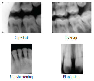
Great care is necessary when placing the X-ray beam at right angles to the dental sensor, to avoid common errors. Incorrectly directing the beam in the horizontal plane will result in overlapping proximal contacts on bite-wing or periapical radiographs, making them diagnostically useless and resulting in a retake.
Similarly, if the X-ray beam is not correctly centered over the receptor, cone cuts can occur on the image, with a clear zone where the X-rays did not expose the sensor. Central ray entry points help to identify the center of the receptor by using an external landmark. In the case of periapical radiographs, improper vertical angulation can produce image foreshortening and elongation that misrepresents the actual length of all structures including the teeth.
Rigid digital x-ray sensors are more difficult to use initially, may result in more errors for both periapical and bite-wing radiographs compared to traditional film, and can cause more discomfort for the patient. To avoid these problems, rigid receptors should be placed close to the midline to aid proper placement and to reduce discomfort. It is particularly important if a patient has a shallow palate or floor of mouth to employ this method, both to avoid discomfort and to avoid distortion of the image.
#4 Distance to the Patient
I see this happening all the time with our customers using our Apex Dental Sensor. The distance between the x-ray head and the sensor can also have an impact on image quality. The further the x-ray head is from the sensor, the lower the amount of radiation is that reaches the sensor. To prevent inconsistent imaging, the x-ray head should be as close as possible to the patient skin. When you are using the holders/positioners for your dental sensor, make sure that you slide the ring on your holder flush with the patients skin. Then make sure your x-ray head tube is flush against the ring. There should be less than an inch gap between the end of the x-ray head tube and the patients skin.
X-ray head generators are a lot like a shot gun. The farther you are away from your target or in your case a dental sensor. The less you are going to hit that target. The closer you are the more likely all of the radiation is going to be hitting the dental sensor.
For every inch of dead space the exposure settings would need to be increased accordingly to achieve the same quality image as if the tube head cone was directly against the patients cheek. A quality dental sensor sensor holder can help ensure your staff are taking the best quality images possible.
#5 Letting go to soon
Dental Sensors can be underexposed if the exposure switch is not activated for the indicated or correct length of time. In other words, the clinician let go of the exposure button too soon. When you set your x-ray generator to a set time say .20 seconds, when you press the button you need to make sure the button is being held down for the duration of that exposure.
I have seen time and time again from doctors wondering why their x-rays are coming out to light, come to find out the are releasing the exposure button to soon.
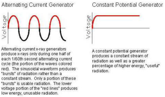
#6 AC VS DC X-Rays
Until relatively recently, almost all dental x-ray generators applied alternating current (AC) to the tube when generating x-rays. Rather than utilizing alternating current, some newer units apply a nearly constant potential to the tube. These units are often referred to as direct current (DC) units. Constant potential generators produce a relatively constant stream of radiation and a greater percentage of higher energy “useful” radiation.
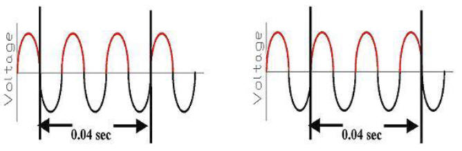
Another consideration occurs at very low exposure times used in digital radiography. AC units may not provide exposures as consistent as constant potential units at these very short exposure times. This property can be illustrated using an example exposure time of 0.04 seconds (which is a very low setting). As stated above, alternating current produces a sinusoidal waveform and x-rays are generated only in the “positive” portion of the waves. A 0.04 second exposure time would cover two and one half 1/60th second alternating current waveforms. Depending on at what point in the waveform the exposure was initiated, as few as two or as many as three “usable” portions of the waves would be captured (at least some, and perhaps all AC units have no control over which segment of the waveform an exposure is initiated). At these very low exposure settings, this could result in a 1/3 difference in exposure for the same 0.04 second timer setting (see diagram below).
Typical AC x-ray generators will typically produce slightly different x-ray each time. It might be a little lighter or darker. To summarize, AC and DC units are both capable of producing diagnostic images whether using conventional film or digital radiography. However, DC x-ray heads will produce a more consistent radiograph.
#7 Time to replace that old x-ray generator
Hate to say it but nothing last for ever. X-ray generators are not exempt from this. Some times they just go bad. The best was to find out if your x-ray generator is going bad is to call the manufacture and get a tech to come look at your unit. Your unit should be serviced everyone in awhile to make sure that it is exposing properly.
When switching from film-based imaging, it is sometimes recommended to refit older X-ray generators with an electronic timer.
Another technical error that occurs occasionally is when the receptor yields no image. This error can be caused by mechanical problems such as electrical failure, faulty generator, timer inaccuracy or faulty exposure switch.
Summary
Intraoral radiographic imaging is an invaluable tool for proper patient care providing critical information for the diagnosis and treatment of dental disease and other oral conditions. To ensure the production of high-quality diagnostic images, the clinician must attend to the principles of accurate image projection when acquiring intraoral radiographic images.
In addition, the clinician must be able to manage the patient effectively during radiographic procedures and be well-versed in the identification and correction of errors when they occur. The clinician is also responsible for eliminating unnecessary retakes and minimizing radiation exposure to the patients under their care.
Weather you are using one of our Apex Dental Sensors or another brand these rules apply. We hope this information helps you not only save time by take less retakes but also allows for you to take amazing radiographs.